Active structures
The term active structures includes muscles. The human body comprises around 650 muscles (of which around 400 are skeletal muscles), which accounts for between 40-50% of a young man's body mass. The muscles can only pull and actively perform movements. Most internal organs as well as the control and regulation systems mainly serve muscle activity. If the muscles are not used, they atrophy. Every year, a person loses around 1% of their muscle mass if nothing is done to prevent it.
The active structures fulfil the following tasks:
posture, statics and movement
damping
metabolism and energy conversion
heat production (body temperature is maintained by muscle activity)
respiration (chest and shoulder muscles serve as auxiliary respiratory muscles)
protection (stabilising the spine and joints and protecting the abdominal organs)
muscle pump (supports blood flow in the veins)
The structure of a muscle
There are three types of muscle tissue:
skeletal muscle
cardiac muscle
smooth muscle fibres in the blood vessels and digestive tract
Structure and function of skeletal muscles
In skeletal muscles, muscle fibres are combined by fine connective tissue sheaths to form muscle fibre bundles. Many muscle fibre bundles form a muscle, which in turn is held together and protected by a coarse connective tissue sheath (fascia). Each muscle fibre contains many myofibrils arranged parallel to the fibre direction.
The myofibrils are tubular, have a diameter of around 1-2 µm and extend along the entire length of the muscle fibre. They consist of sarcomeres arranged one behind the other. This is why they appear striated under the microscope.The sarcomeres are the contractile functional units of the myofibrils. They consist of a series of protein molecules connected in series, the actin and myosin filament. The actin and myosin filaments overlap so that the myosin heads can interact with the actin filaments. When the muscle is activated by the nervous system, this interaction leads to a contraction of the muscle. In a contraction, the myosin heads pull the actin filaments together; this happens simultaneously throughout the muscle, which then leads to the contraction of the entire muscle. This process requires energy in the form of ATP.


Fibre types
There are different types of fibres that make up the muscles. There are roughly two types:
Type I fibres
ST fibres (slow-twitch) or dark or red fibres are rather thin, slow-twitch muscle fibres and therefore tend to contract slowly. They are designed for continuous performance (e.g. marathons, cycling tours) with limited effort and only fatigue very slowly. Accordingly, they are mainly found in muscles that fulfil long-lasting static tasks. The ST fibres obtain their energy aerobically, whereby the oxygen required for this is taken from the blood. They are also known as type 1 fibres and oxidative fibres.
Type II fibres
FT fibres (fast-twitch), also known as light or white fibres, are thick and fast-contracting muscle fibres. They consume more energy and fatigue more quickly. They are also called type II fibres or glycolytic fibres. Muscles that fulfil dynamic tasks (e.g. sprinting, boxing) are made up of these fibres. There are two further groups of type II fibres: type IIa fibres (FTO fibres) and type IIb fibres (FTG fibres). The FTO fibres are relatively more resistant to fatigue than the FTG fibres, which can contract more quickly.
The distribution of muscle fibres is largely genetically determined. The conversion of FT fibres into ST fibres is possible, but the reverse is hardly feasible (everyone can become faster, you are born a sprinter). The intermediate type (FTO) is located between red and white muscles in terms of its characteristics. In contrast to the others, this type can be influenced in one direction or the other through targeted training.
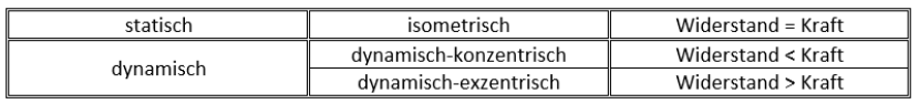
Forms of contraction
Muscle work means contraction of muscles. Not every movement requires the same type of contraction, so a distinction is made between two forms.
Muscle groups


Each joint is controlled by (at least) two muscles, which act against each other. The agonists and antagonists are equal partners. They take on the leading role alternately. Only their complementary action enables coordinated movements. The agonists do the main work. As counterparts, the antagonists control the movements by slowing down, dosing and stabilising. When agonists and antagonists are equally (statically) active, their torques cancel each other out so that certain joint angles and postures are fixed.
Stabilising muscles
These muscles consist mainly of the enduring ST fibres and can therefore perform light work over a longer period of time. These muscles are responsible for stabilising the spine, the shoulder joint, the shoulder blade and the arch of the foot. The development of these stabilising muscles is of great importance for a healthy posture. If the stabilising muscles are not sufficiently developed, this can lead to overuse of the locomotor muscles because the lack of stabilisation is compensated for by the use of larger muscles. However, the locomotor muscles are not designed for such prolonged strain.
Movement muscles
These muscles are responsible for so-called target motor skills, i.e. all dynamic tasks. They consist more of the FT fibres. The locomotor muscles are used to perform all heavy work that is not performed for long periods of time. As described above, it becomes a problem when movement muscles are used to compensate for weaknesses in the stabilising muscles, which is why it is very important that both muscle groups are trained equally well.
More info:
Training explained - Handbook of training theory (Jost Hegner):
The active part of the movement and support system