Passive structures
The term passive structure covers all parts of the musculoskeletal system that cannot move on their own, i.e. cannot move actively.
Bones

Depending on the source, the skeleton consists of 206-212 bones, which can range in size from a few centimetres to half a metre. It is constructed according to the principle of lightweight construction, which means great strength and resilience with as little material as possible (approx. 7 kg).
Construction and function are closely related:
Long bones:
favourable levers and movement (arms, legs)
Short bones:
resilient load-bearing function (spine, foot, hand)
Plate bones:
support and protection (skull, ribcage, pelvis)

Build-up
The bones consist of a compact "cortex" and a system of fine bone beads. These are aligned in such a way that the forces occurring are optimally absorbed. Each bone is covered by a rough connective tissue membrane, the periosteum. This protects the bone and is rich in blood vessels and pain receptors.
Length growth
The cartilaginous growth zones (epiphyseal joints) do not ossify until longitudinal growth is complete. There is therefore a risk of injury and permanent damage due to excessive strain in adolescents.
Osteoporosis
Increased bone fragility is a disease of civilisation that can occur in older people. Effective prevention involves building up a large bone mass through plenty of exercise, sport, a healthy diet and avoiding nicotine.
Joints
The bones are connected to each other by joints. The different types of joints enable the body to move:

Hinge joints:
movement around an axis (elbow joint, finger joint)
Saddle joints:
movement around two axes (metacarpophalangeal joint of the thumb)
Ball and socket joints:
movement around three axes (hip, shoulder joint)
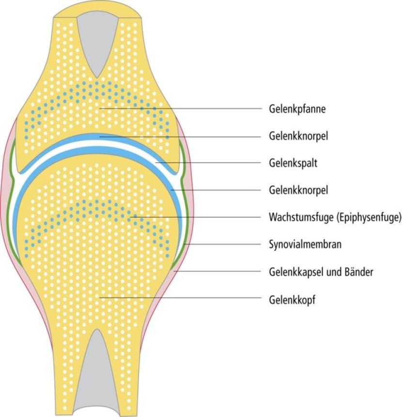
Each joint consists of a joint socket and a joint head. Between the mirror-smooth joint surfaces made of hyaline cartilage is a wafer-thin space, the joint space. The joints are stabilised and secured by ligaments (passive) and muscles (active). Each joint is enclosed by a joint capsule made of coarse connective tissue. This is lined with a fine epithelial tissue, the synovial membrane.
Cartilage
The joint surfaces are covered by a layer of cartilage without blood vessels and kept moist by a mucous fluid, the synovial fluid. This ensures a high level of compressive strength and enables resistance to sharp forces. Bruises, sprains and dislocations damage this tissue and, if repeated or treated improperly, can lead to permanent joint changes (arthrosis). In the knee joint, two crescent-shaped wedges of cartilage, the menisci, enlarge the flat socket of the tibia. Meniscus injuries (tears, bruises) often occur when skiing, playing football or handball.
There are three forms of cartilage:
Fibrous cartilage (intervertebral discs, pubic symphysis, menisci in the knee joint)
Elastic cartilage (scaffolding for nose and ears)
Hyaline cartilage (elastic contact, gliding and protective layer on joint surfaces)
Hyaline cartilage is not supplied by blood vessels, but is nourished by the body via synovial fluid (synovial fluid). As a result, the metabolic activity is very low, so that the cartilage can hardly repair itself or adapt to higher loads. The cartilage is not sensitive to pain, which is why wear and tear or injuries cannot be recognised by the senses. The water content in hyaline cartilage tissue decreases over the years, which means that cartilage and joint injuries can occur more quickly with age. Mobilisation exercises can stimulate the release of synovial fluid and thus prepare the cartilage for the upcoming load.
Spine

The spine is made up of a large number of vertebrae, which are connected to each other by small joints and cartilaginous intervertebral discs. The intervertebral discs, which are located between the bony vertebrae, provide good cushioning against shocks and sharp forces.
The spine is curved in an S-shape and is passively stabilised by ligaments and actively stabilised by the muscles. The most important functions of the spine are to protect the spinal cord (and nerve cords) and to stabilise the trunk. Torso stability is an important prerequisite for spinal health, quality of life and any sporting performance. It depends primarily on the strength and commitment of the trunk muscles.
Tendons and fasciae, ligaments, tendon sheaths and bursae
Tendons and fasciae, tendon sheaths and bursae
All muscles are attached to the skeleton by tendons and are used to transmit force. The tendons consist of tight collagenous connective tissue. To ensure that the tendons move with little friction and remain attached to the bone or ligaments in an optimal position, they run in tendon sheaths. These are attached to the bone or ligaments and are filled with synovial fluid, which reduces frictional resistance. Where tendons run over bones, bursae filled with synovial fluid serve as cushioning. Tendons adapt to changes in load by the connective tissue fibres becoming more numerous, thicker and more resilient. We can distinguish between tendons of origin and attachment tendons. The tendon that lies closer to the centre of the body is called the tendon of origin, the other is the tendon of insertion.
Fasciae are a type of muscle skin that cover individual muscles or entire muscle groups.
Ligaments
Ligaments connect bones across joints. They are made of the same material as tendons and can also adapt to changes in strain.
When you move your foot in all possible directions, you realise that the movements are only possible up to a certain point. If the foot were to be forced beyond this point, the joint would be dislocated. However, before this could happen, the ligaments would first be strained, overstretched or even torn. The ligaments therefore prevent excessive swings in movement, they guide, support and hold, but are not very stretchable and can hardly be trained, although they do have a good ability to regenerate (e.g. after a tear).
Weak points in the skeleton
The quality of the connective and supporting tissue depends on predisposition, age, training status, hormonal balance, diet and the bone mass and density built up during adolescence. There are major individual differences in terms of exercise tolerance. When moving, the following weak points should generally be taken into account, which can quickly be damaged by incorrect posture in everyday life or overuse in sport:
Spine;
Growth joints of the long bones (not yet closed in children and adolescents);
Hyaline cartilage of the joints (cartilage is not sensitive to pain, so injuries often go unnoticed);
Tendons, ligaments, tendon sheaths and bursae.
More info:
Training fundiert erklärt – Handbuch der Trainingslehre (Jost Hegner):
Der passive Teil des Bewegungs- und Stützsystems